How to Take an In-Brace X-Ray
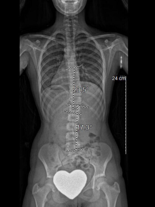
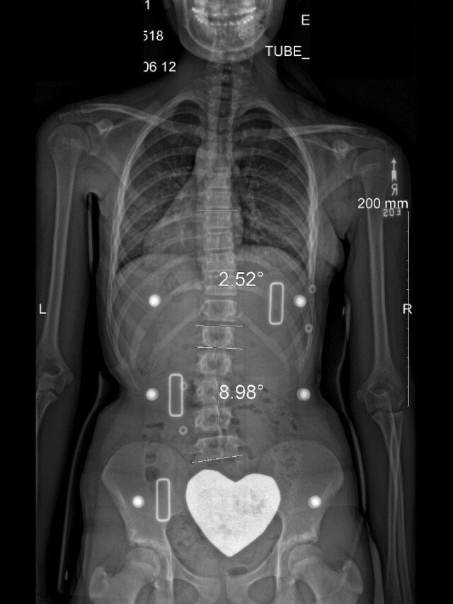
In-Brace X-Rays are critical for evaluating the effectiveness of any scoliosis orthosis. These X-Rays are the primary tool for determining the impact the brace is having on the patient internally, and they help to inform the Orthotist of any adjustments or modifications that might be needed to improve correction and outcomes. Being able to review In-Brace X-Rays, regardless of the brace’s style, is important in evaluating mold modifications and ensuring effective brace blueprint design.
This will also help our team of scoliosis specialists successfully maintain the highest quality for you (the clinician), your scoliosis referrals, and, most importantly, your patient.
Full-Time In-Brace X-Ray Protocol
It is critical that your In-Brace X-Ray is accurate. Download a PDF to bring to the radiologist to help ensure best results.
Important Note:
To properly evaluate the design and effectiveness of the orthosis:
- The patient must be wearing the brace positioned on their body exactly as their
orthotist has instructed them to do. - The brace must be worn at full tightness.
- Providence® Brace X-Rays must be taken with the patient lying down in supine position.
- Full-time braces (Boston, Chêneau, Wilmington, etc.) X-Rays must be taken standing.
Providence® In-Brace X-Ray Protocol
It is critical that your In-Brace X-Ray is accurate. Download a PDF to bring to the radiologist to help ensure best results.


