Principles of 3D Bracing – Design and Methodologies
Orthotic Management of Adolescent Idiopathic Scoliosis (AIS)
Section 4
Three dimensional bracing is not a new concept by any means. Dating as far back as the late 5th century, when Hippocrates identified Scoliosis (root “skol”: meaning twists and turns), it has been well understood that the nature of the deformity presents in multiple planes. Orthotic approaches to managing these characteristics have long taken into account the dynamics of de-rotation, medial translation, and balance. Much of what you will find in 3D bracing is achievable in a well-adjusted, modified, and custom fit orthosis that began as a symmetrical module, albeit quite a bit of time and effort to get there.
Related Links
Need more information?
Contact our Customer Service Team at 800 253 7868. We'll be glad to help you with your question.

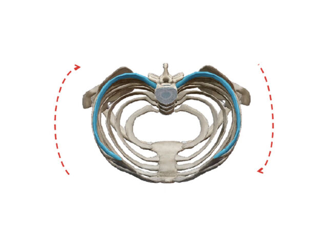
3D Bracing incorporates the de-rotational and translation forces applied through the ribs
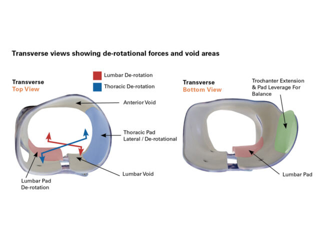
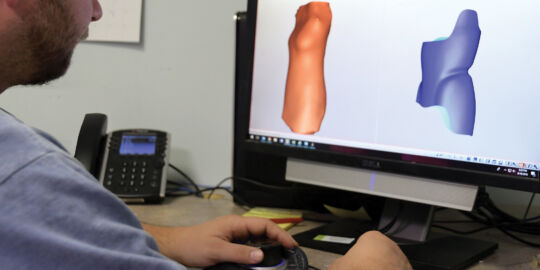
However, the methods, tools, and accuracy have changed with the incorporation of scanning and CAD design. This is where we begin to see the value of what has been termed “3D Bracing”. The advantages arise from direct manipulation of the patient model in a digital format. Rather than adding localized pads to generate our forces, and heating or cutting out spaces for reliefs, the same can be done effectively by de-rotating and shifting segments of the patient’s model as a whole. Any segmental changes made will inherently create forces and reliefs, as the section is moved as a complete unit. This becomes most beneficial when addressing the Thoracic region. Rather than having a singular pad contributing to the de-rotation and translation forces transferred through the ribs on one side, capturing the entirety of that segment and manipulating it as a whole greatly improves accuracy, increases leverage, and adds more comfort for the patient by maintaining a greater surface area for contact.
By working directly with the patient model, form-fitting contact can be maintained in most areas, while providing for ample relief where necessary for corrective movement. In addition, modification of the model prior to brace fabrication offers the ability to incorporate some features not easily created after the fact, when the brace is being fit. The best examples of that can be seen in the anterior de-rotational void in front of the posterior-lateral thoracic force, as well as the anterior thoracic de-rotation force on the contralateral side and the accompanied posterior relief.


